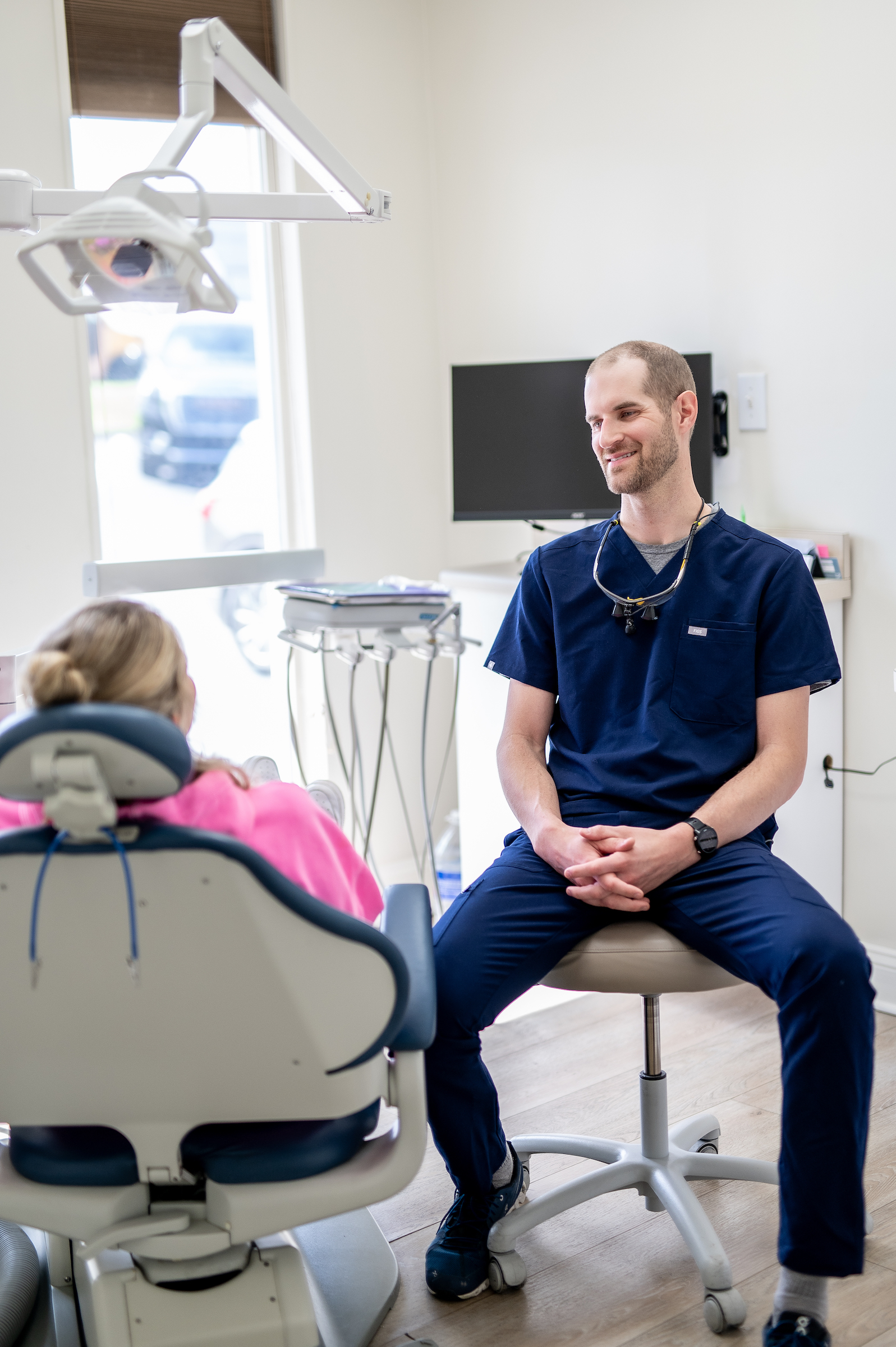


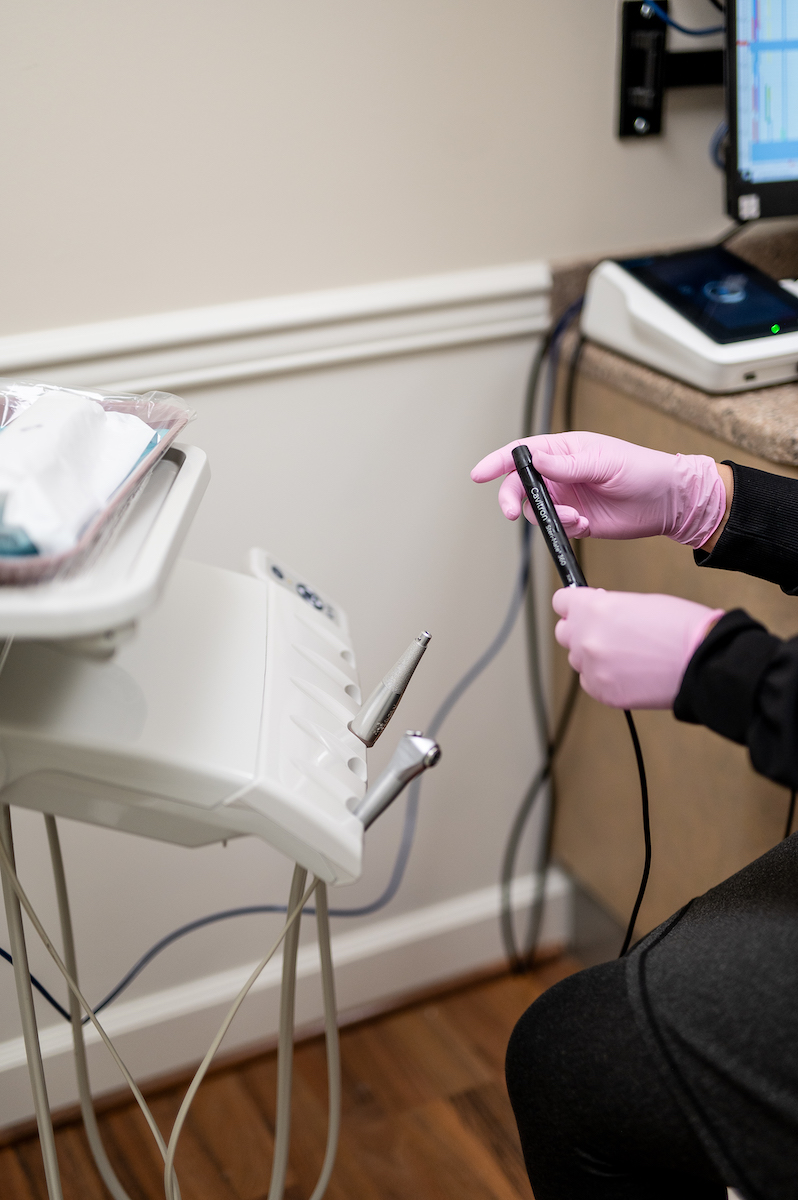
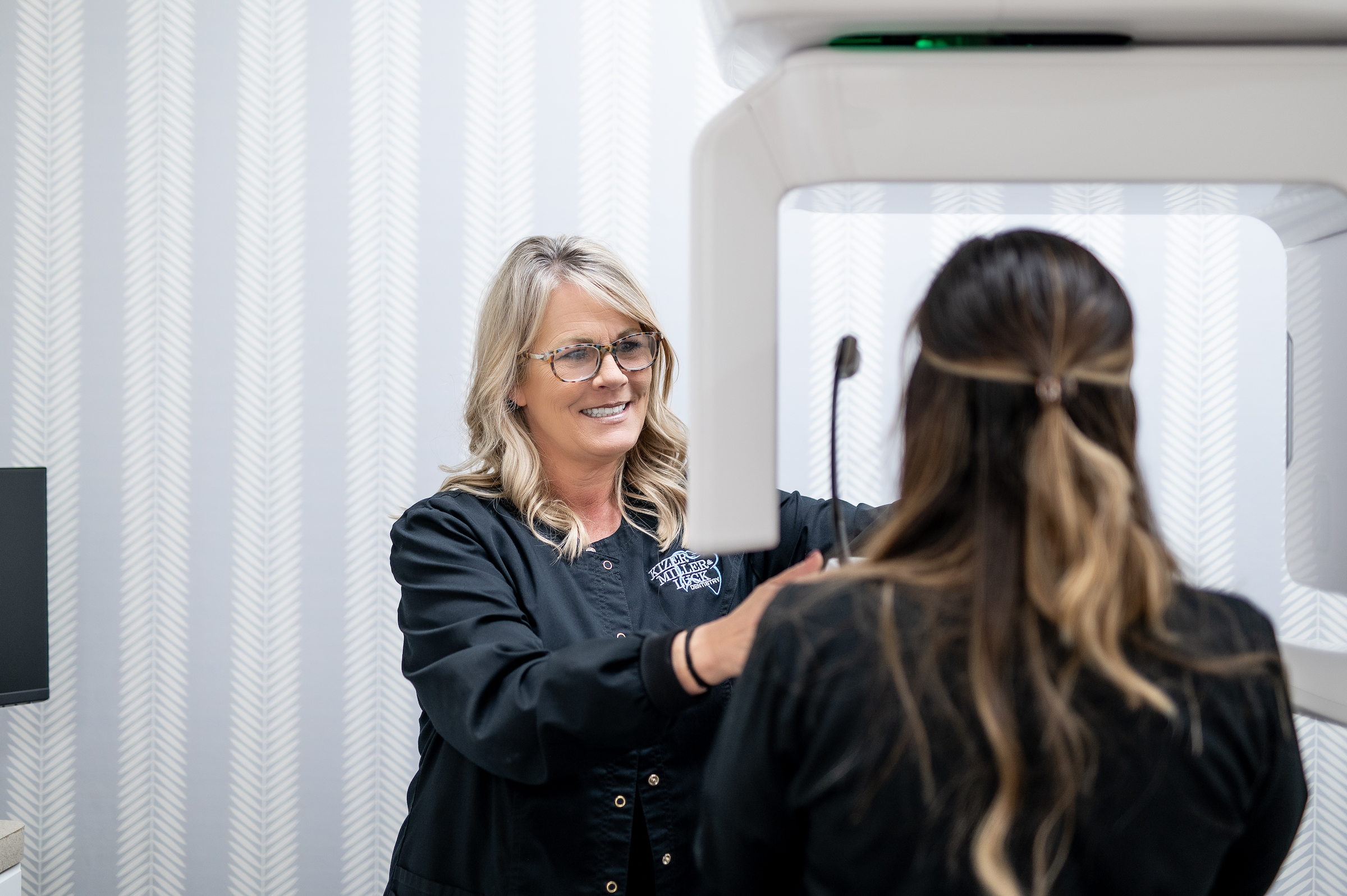
CBCT: Cone Beam Computed Tomography is a revolutionary imaging technology that allows our doctors to see the anatomy of the upper and lower jaws in three dimensions. This type of image shows far more diagnostic information than a typical panorama or bitewing x-ray. It is used to diagnose different types of pathology, fractured teeth, sinus and nasal cavities, planning for precise implant placement, and the location of certain vital anatomical structures.
Intraoral scanning: Digital intraoral scanning has virtually eliminated the need for the messy impression materials used in the past. Our dentists and assistants use a scanning wand to digitally stitch together a “digital impression” of your teeth with proper bite alignment to ensure a highly accurate and mess-free digital copy of your teeth for various procedures.
Intraoral Camera: A picture is worth a thousand words. Intraoral cameras have aided the dental professional in showing different dental diagnoses. It’s one thing for your dentist to say that you need a filling or a crown, but when you have access to the full view of the tooth to understand the diagnosis, it helps to understand exactly why the treatment is recommended. We use images from the intraoral camera to better describe the prescribed dental treatment during your hygiene and dentist visit.
Guided Surgery: Guided implant surgery significantly reduces the uncertainty associated with the placement of dental implants. In combination with the CBCT and intraoral scan, our doctors often virtually plan the placement of your dental implant before you even arrive for surgery day. Guided surgery allows for accurate angulation, depth, and position of the implant which leads to more precise restorations and gum health around the implant.
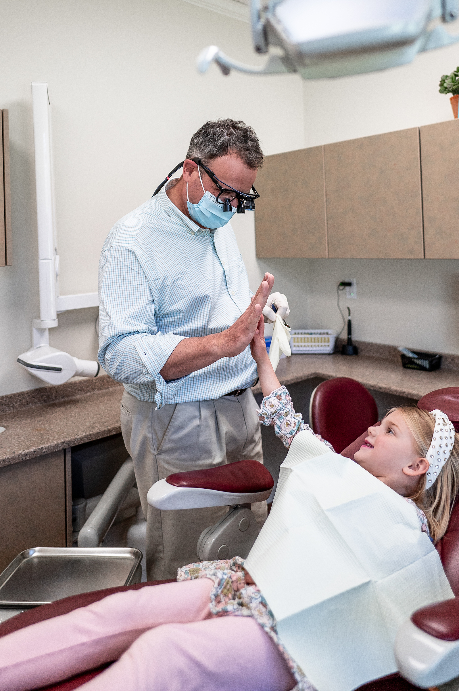
The Kizer, Miller & McKnight crew are always happy to see you 🪥

Tooth be told, you are awesome! 🦷 we have the best patients ever! Look what we got from one of the best! #grateful

Let’s hear it for these beautiful ladies 🪥 our incredible dental assistants make our world go round at KMM! @kizermillermcknightdentistry

It’s a good day for a cleaning 💦 @kizermillermcknightdentistry
#kizermillermcknight #knoxville #dental #dentist

Welcome to KMM!
We are so glad you’re here! ☀️
#dentistry #knoxville #dentist #dental #865life #dentistlife #farragut #bearden #middlebrook #smile

The gangs all here and ready to take on a new week making your teeth shine ✨ @kizermillermcknightdentistry
@thelenwrightphotography

The best dental care in Knoxville with two locations!

It’s a good day to visit Kizer, Miller & McKnight Dentistry ☀️ #goodmorning

Everyone loves party favors 🎈 coming to see us and leaving with these goodies is basically the same thing #brush #floss #smile

New year, same amazing team… Kick off the year with us! We can’t wait to see you at Kizer, Miller & McKnight Dentistry

